Posted on

What is Mobile Imaging?

Medical imaging has been revolutionized by the invention of mobile imaging. Mobile Xray imaging is a new way of delivering medical imaging services from anywhere, at any time, and under any conditions. It combines the best benefits of traditional medical imaging with an unparalleled level of convenience and accessibility.

If you need to find out more about mobile imaging, just keep on reading this article because we’ll be explaining everything.

What is Mobile Imaging?

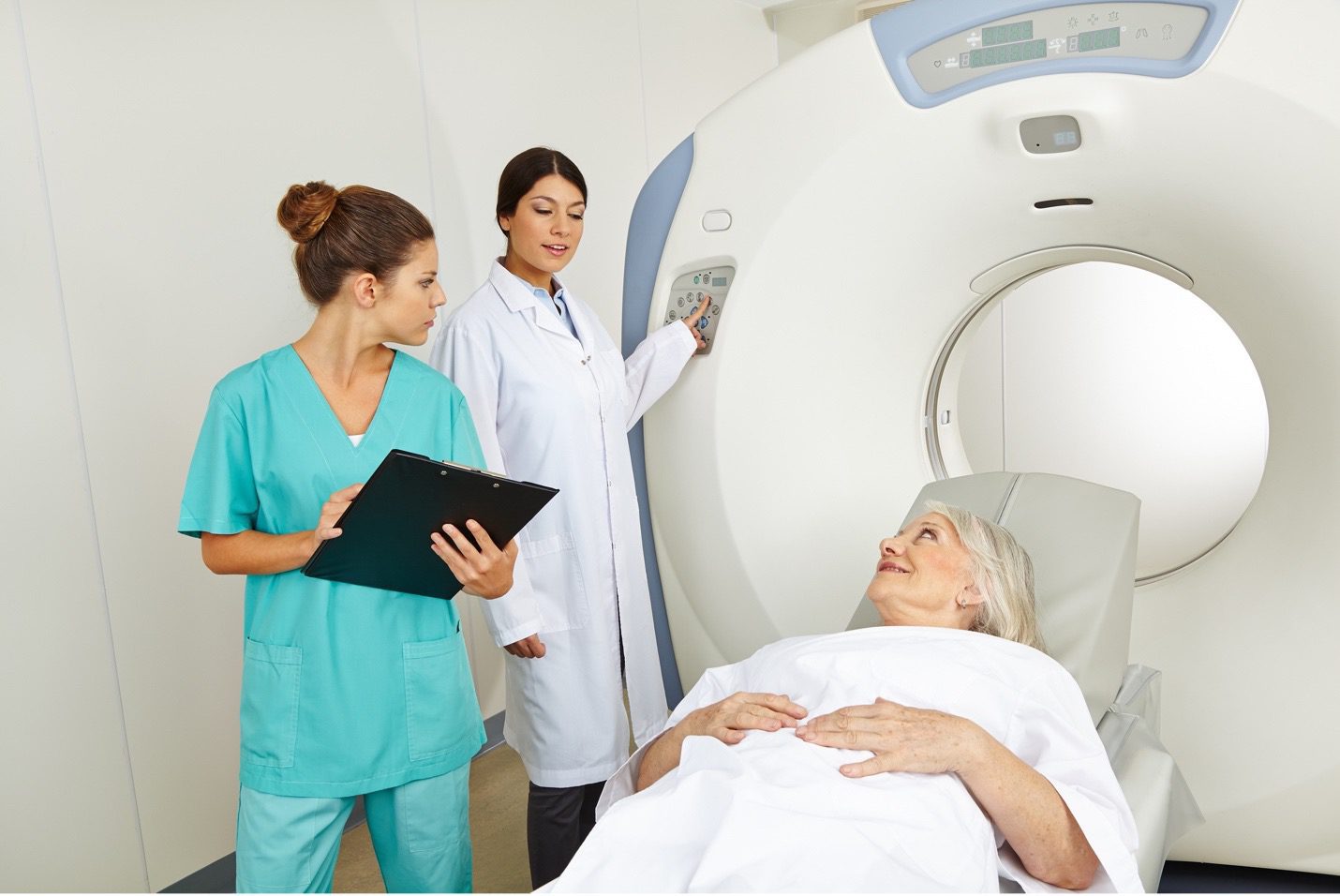
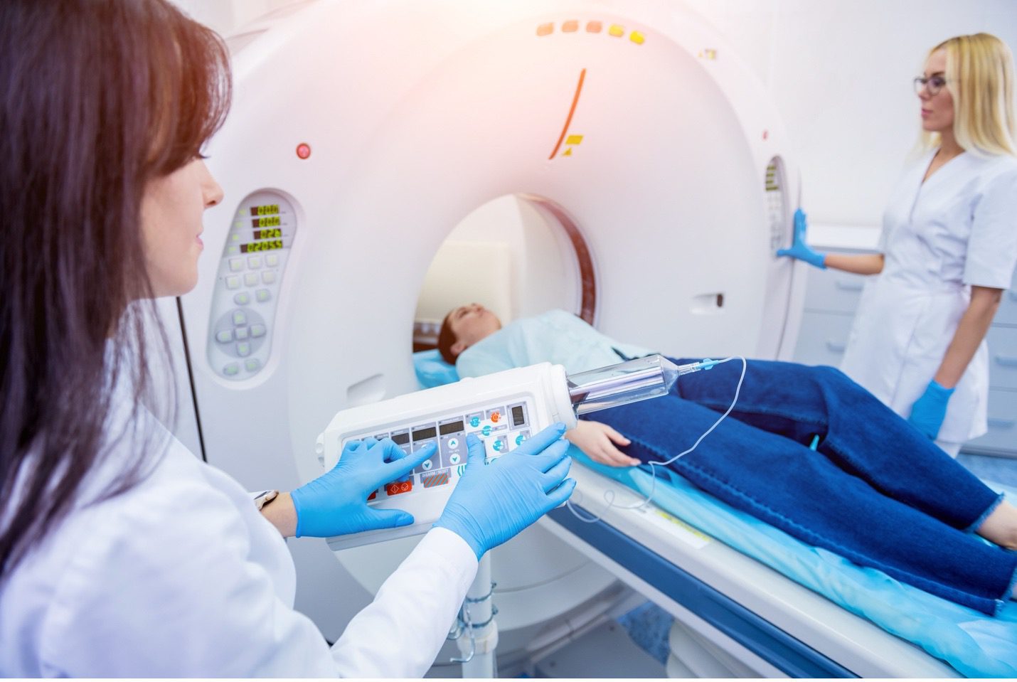
Mobile medical imaging is a service that provides medical imaging services via the use of mobile radiology equipment. These portable imaging devices are designed to provide diagnostic services in the most convenient and accessible settings.

Mobile imaging systems have been developed over the years as an alternative to fixed radiology departments. It’s faster, more convenient, and less expensive than traditional hospital-based imaging services. The mobile service allows patients to get their tests done anywhere, at any time.

The most common types of mobile radiography machines are Xray, EKG, and Ultrasound machines, among others. These machines come with high-quality functions that ensure accurate results every time they are used by certified technologists and radiologists. They can be used for various purposes:
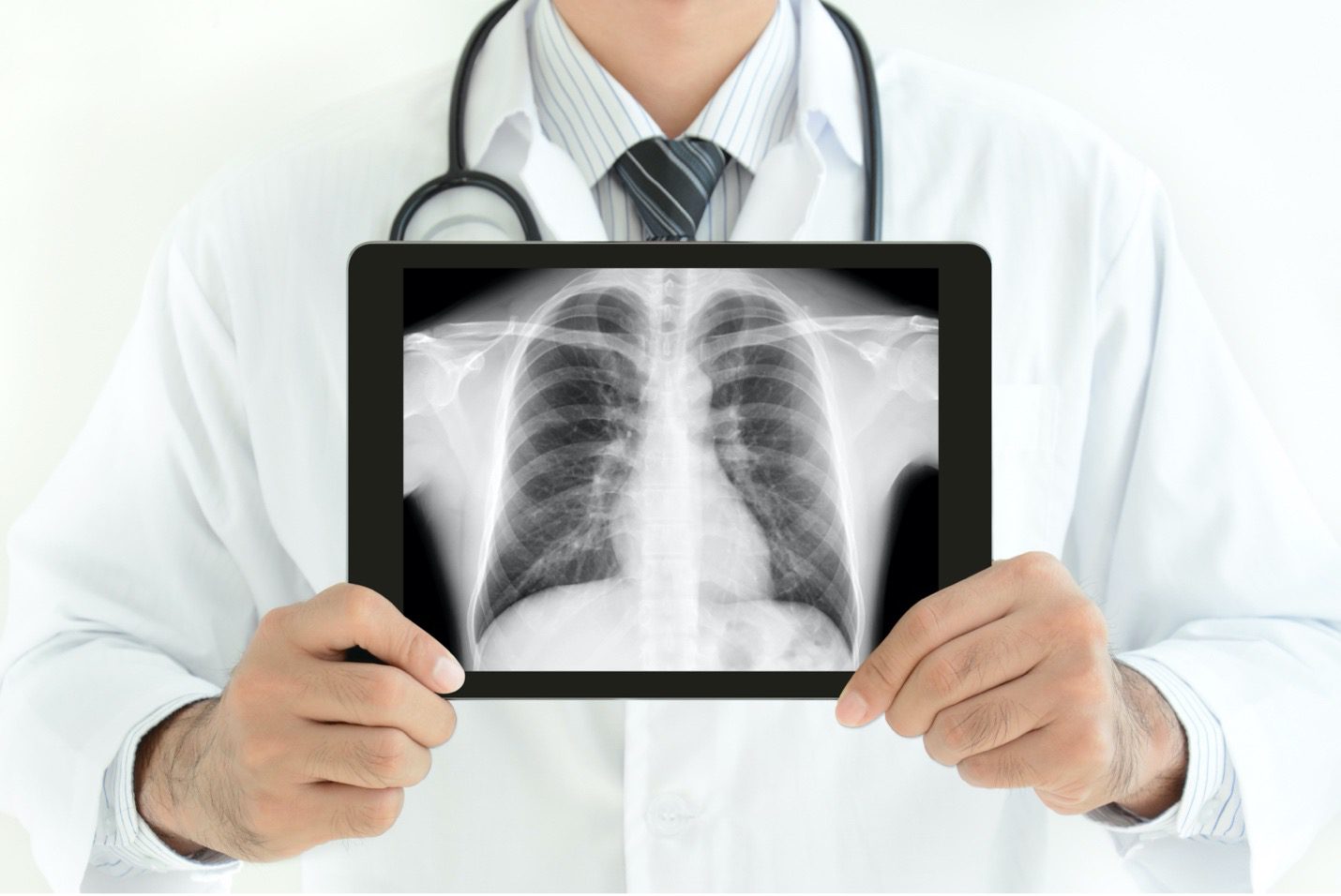
Xray

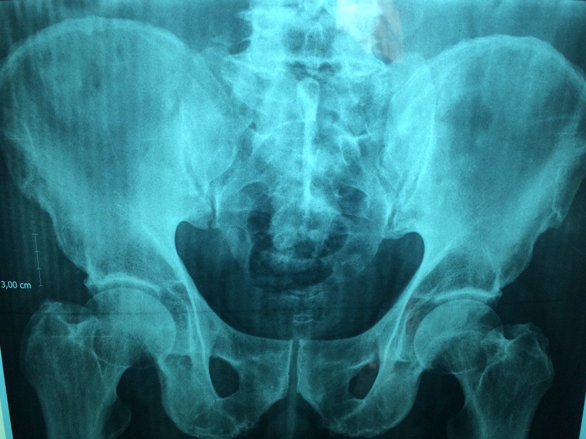
Xrays are a type of noninvasive medical imaging technique that uses ionizing radiation (radioactivity) to produce images of internal tissues, organs, and bones. They are most commonly used to diagnose bone fractures, injuries, deformities, and other conditions that involve broken bones. They can also be used to identify whether an implantable pump or catheter is properly placed in the body and whether pneumonia or bronchitis has developed inside the lungs.

EKG

The EKG is a test that checks whether your heart is healthy or not. It measures the electrical activity of your heart and makes sure that it’s working properly. It can detect problems in the heart itself, such as an irregular heartbeat, which can be caused by a variety of things: high blood pressure, high cholesterol levels, or even coronary artery disease (narrowing of the arteries).
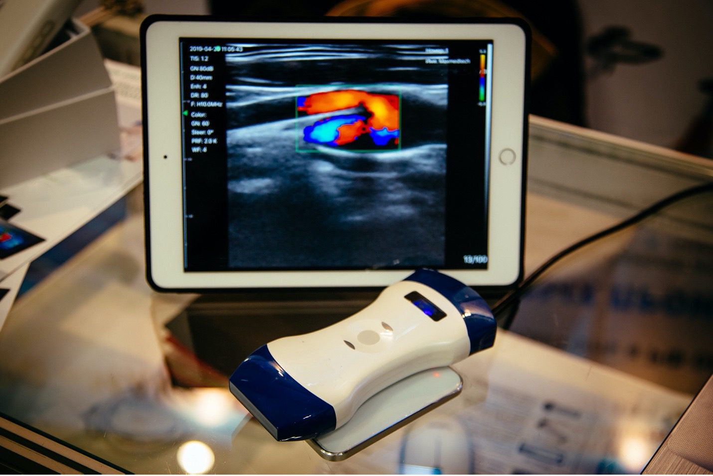
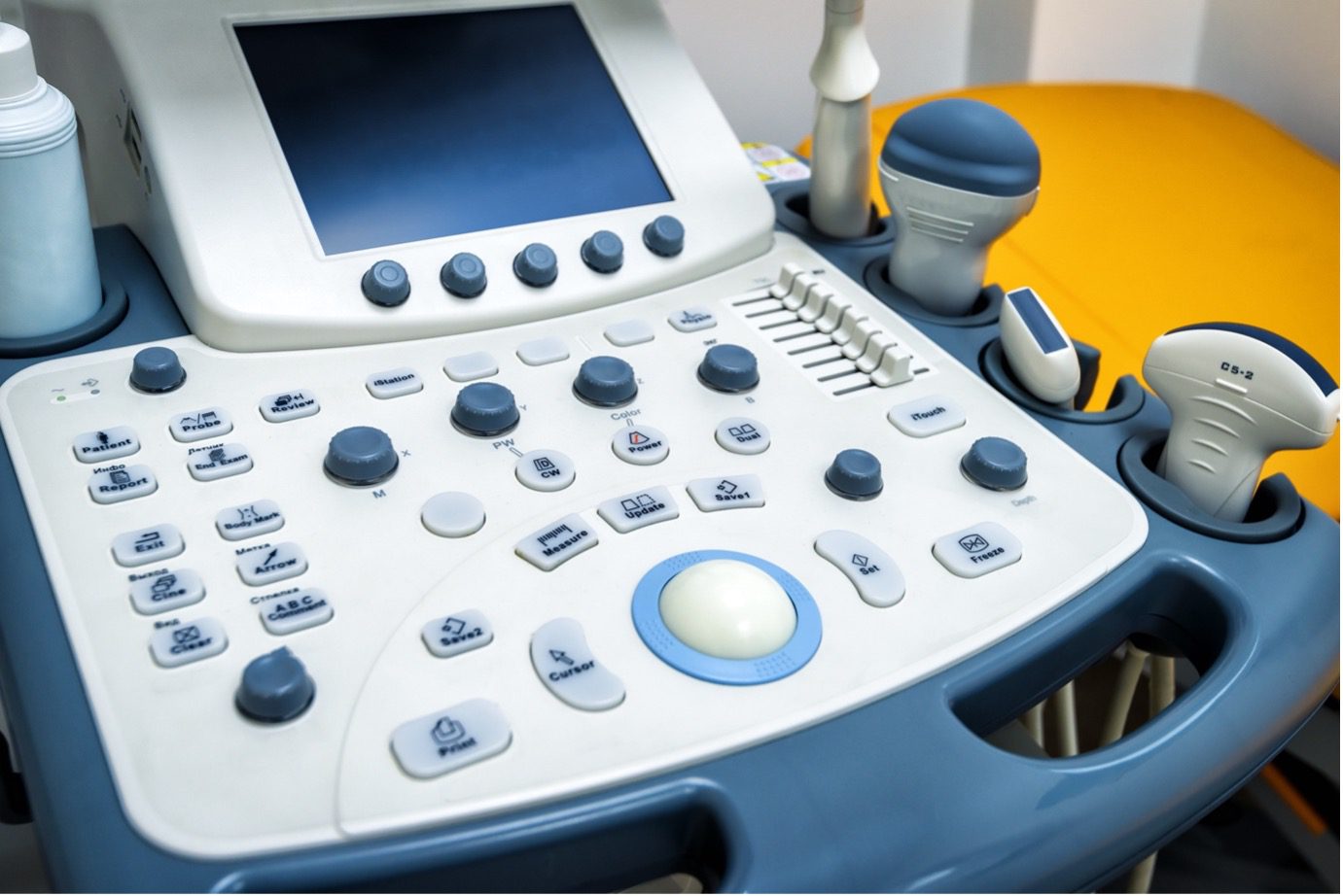
Ultrasound

Ultrasounds are a type of imaging technology that uses sound waves to take pictures of internal structures like organs, bones, and muscles. The ultrasound images can show abnormalities in these structures and be used to diagnose diseases such as abdominal aneurysms, blood flow issues in carotid arteries (which carry blood away from the heart), renal vascular disease (which causes abnormal clots), signs of kidney failure, or other conditions.

Benefits of Mobile Imaging

Mobile medical imaging has many benefits for patients and healthcare professionals alike. The reasons why people prefer to use mobile units include:

Affordability – compared with going to a clinic for an Xray or ultrasound scan, most people will find that mobile imaging is more affordable.

Quick result – it takes less time to get your results than if you went to the hospital for a traditional appointment.

Convenience – you don’t need to wait for an appointment at the clinic for your screening tests; you can make appointments online or by phone anytime.

More versatile – mobile equipment is more versatile than fixed units, which means that it can be moved around and used wherever it is most convenient for the user.

Conclusion

This new technology is making it possible for patients to have their imaging tests done at home, at work, or in the comfort of their own vehicle. It has become more accessible, less expensive, and efficient to those who need it most. As the mobile  Xray and medical imaging industry develops, expect to see much larger networks and more efficient services in the near future. To learn more about mobile imaging or to inquire about what mobile imaging equipment we offer, contact us today!

Posted on

Best Chiropractic Tables

There is no one-size-fits-all answer to this question, as the best chiropractic tables will vary depending on your needs and preferences.

Primary Considerations

The type of table that you choose will largely depend on your body composition, height, and build.

2×4 Tables

This style is typically used by practitioners who work with people who are taller than average or those who have a larger frame, because it allows them to adjust patients more easily from a standing position.

3×3 Tables

This style is often preferred by practitioners who work primarily with lower-body injuries because it allows them to provide treatment from multiple angles simultaneously without having to move their patients excessively.

4×2 Tables

These tables (also referred to as double tables) allow practitioners to treat both upper-and lower-body problems at once, since they’re wider than standard 2 x 4s but shorter than 3 x 3s.

Most Popular Types of Chiropractic Tables

There are several types and models available on the market today, so it’s hard to decide which is the best for your practice.

  • Manual – These are the traditional type of chiropractic table that uses force to adjust your spine. They are often used by chiropractors who specialize in neck pain relief or back pain relief.
  • Adjustable – These tables can be adjusted to provide the best alignment for your spine.
  • Magnetic – These tables use magnets to adjust and stabilize your spine, providing relief from pain and improved function.
  • Elevation – Chiropractors use elevation tables to place patients at the right height for maximum comfort and best results.
  • Hi-lo – This table can be used to raise patients vertically. This table is especially helpful for patients who are less mobile.
  • Stationary – A low maintenance table with few moving parts. Although they may not offer all the benefits of more complicated tables, stationary tables are less likely to break or malfunction.
  • Decompression – Designed primarily for accident victims or patients with bulging, degenerating or herniated discs, these tables lengthen the spine to reduce pain in the back and leg areas.

Best Chiropractic Tables

Elite chiropractic table

This table can be adjusted to suit any patient’s body type and position. If you are a chiropractor, this table is ideal for you. You can adjust it to accommodate different patients and different degrees of adjustment. The table has no screws or bolts to come loose and get lost, so you can be sure that this table will be here for years to come.

The Flexion Table

The Flexion table is designed for use by chiropractors who specialize in spinal manipulation and flexion adjustments. It is versatile and can be adjusted to accommodate a wide range of patients and treatment scenarios.

Qualities to Look for in a Chiropractor Table

When looking for a chiropractic table:

  • Make sure that the table is sturdy enough to support your weight and has adjustable height options.
  • Look for a table with casters so that you can move it around easily.
  • Ensure that the surface is smooth so that your patients don’t experience discomfort while you are treating their neck or spine.

Call us at Great Lake Imaging to learn more.

Posted on

Medical Imaging for Women

Women need special treatment and testing at every stage of their lives. Medical professionals can use these tests to quickly and accurately diagnose their patients.

Some medical equipment suppliers specialize in providing the latest screening technology.

Mammogram

A mammogram is a series of x-rays taken of the breasts.

These images can help doctors detect early signs of breast carcinoma, such as abnormal breast tissue changes or lumps that are too small to feel. Early detection and treatment often make it possible to prevent further complications.

Technologists conduct annual mammograms in order to detect any undiagnosed breast cancer. This screening helps to prevent breast cancer and provides peace of mind.

Diagnostic mammography

If a screening exam shows any signs or symptoms, a doctor may recommend that a patient undergo a diagnostic mammogram. If abnormal symptoms, such as skin dimpling or nipple drainage, the patient may be referred for further diagnostic testing.

Ultrasound

Ultrasounds are high-frequency sound waves that give real-time images of the patient’s body. The ultrasound test can provide accurate images because sound waves travel through fluids and soft tissues, but bounce back when they touch denser surfaces. Because it does not use radiation, it is safe for pregnant women.

Ultrasounds are also used by doctors to detect cervical and ovarian cancer.

Follow-ups on mammograms: If the woman’s mammogram is not complete, the doctor might recommend an ultrasound to examine breast tissue abnormalities or other conditions.

Breast MRI Scans

If an ultrasound or mammogram test does not provide enough information to make a definitive diagnosis, a breast MRI will be recommended by medical professionals. If they require more information, they may perform fetal MRI scans on pregnant women.

Breast MRI scans use radiation to create a detailed image by scanning the breast from different angles. This allows doctors to assess tissue type and density.

Patients at higher risk for breast cancer can have a breast MRI done as a screening tool. This diagnostic test is not meant to replace a mammogram, ultrasound, or other imaging tool. It serves as an additional tool for accurate diagnosis and treatment.

Bone Density Scans

A bone density scan is used by medical professionals to determine the amount of calcium per sq. centimeter in a patient’s bones. Doctors can use this scan to determine the likelihood of fracture in patients and to treat those with lower bone density or lack of minerals.

Women are more susceptible to osteoporosis than men because they have smaller bones than men. Their bone density should be checked as their estrogen levels drop during menopause. This test can determine a patient’s bone fragility, and the best ways to avoid fractures.

DEXA

For a complete body composition scan, a doctor will recommend that a BodyLogic(TM), DEXA Body Composition scan be performed. This will allow for detailed images of the patient’s entire body. Medical professionals can use this information to accurately assess the patient’s health and provide the best treatment plan. The doctor can immediately analyze the patient’s health with a DEXA scan.

Visit GreatLakesImaging.com to learn more about medical imaging for women.

Posted on

Proper Setup of Medical Equipment

A competent, skilled, and experienced staff is the backbone of any hospital or other healthcare facility. However, exceptional patient care also depends on having up-to-date functional technology.

Using technologically advanced equipment improves patient satisfaction and increases staff efficiency, which leads to higher revenue. How do you make sure that your new medical equipment is ready to use immediately after you’ve ordered it?

Here are five tips to ensure the most efficient and smoothest equipment setup.

Inspection of Parts and Proper Assembly

Source / On-site Supplier Inspection

Source inspections take place at the supplier before shipment to your facility. The source inspection is the first step of the process. It checks for nonconformance and defects and determines the production order quantity.

These supplier inspections verify compliance with equipment plans prior to production. Depending on the level of delegation, source inspections can either be done by an internal or external provider.

Receive Inspection

A receiving inspection acts as a gatekeeper before a product is accepted. At a specific stage in the product’s lifecycle, received materials, components and finished goods are checked. Inspectors are responsible for ensuring that equipment arrives in proper working condition. They also need to evaluate the material for damage in order to avoid future problems.

For the following reasons, you should carefully inspect every piece of equipment:

  • Missing Parts
  • Damaged Parts
  • Incomplete order

This is an important step for any hospital or healthcare facility, even though it might seem obvious. To avoid future problems, a quick inspection is not enough.

It doesn’t matter how well you put together medical equipment. After you have inspected all the pieces, make sure everything is assembled correctly. A small mistake could cause equipment malfunctioning and potentially damaging your equipment. This can lead to more money and time spent on equipment.

Select the location for the equipment

A majority of offices have a basic diagnostic system. It includes the instruments necessary for physical diagnoses, such as an otoscope and ophthalmoscope. These systems can be mounted on the wall to make them easy to access. You and your staff will not have to change your routine every time you enter a new room if the systems are mounted in the same location in each exam room.

You need to have the equipment located in the area where it will be used, so a direct-to-site delivery process is extremely beneficial.

Test of Functionality

After assembly, it is important to test the equipment for functionality. Even though everything may look perfect, there could be a problem that cannot be seen. Testing can reveal assembly errors before permanent damage is done.

Instruction for Use

Finally, after all inspections have been done and everything seems to work properly, it’s crucial that each applicable member of your staff is instructed on the proper use of the medical equipment, especially if it’s a new model that’s not been used by your staff.

Clear, detailed instructions prevent misuse, which could lead to poor patient care or damage of the equipment. It’s also important that instruction includes the hospital biomedical staff in order to facilitate shock-free operation and proper electrical grounding.

Great Lakes Imaging can set up your medical equipment for you. Call us to learn more.

Posted on

The Importance of X-Rays in Veterinary Care

new and used veterinary equipment

X-rays for veterinarians are among the oldest, yet most frequently used diagnostic tools for medical imaging. Dating back to its invention in the late 1800s and as early as 1939, it was recognized as a revolutionary tool for animal use.

The X-ray machine has become an essential piece of veterinarian equipment due to its continual advancement as a diagnostic tool. It is often one of the first things to be used in many clinics. The turn toward digital imaging has made it even easier for a clinic of any size to have an image read by a certified radiologist within minutes.

How X-Rays Improve Veterinary Outcome

An X-ray by a veterinarian improves patient outcomes because of the ability to diagnose an animal quickly. In the past, veterinarians palpated the animal, drew blood, or obtained urine samples to determine if something was wrong with the animal. While veterinarians can still use these procedures today, time can be saved; a quick, painless X-ray can diagnose the problem accurately.

Earlier, veterinarians used the X-ray primarily to determine a broken bone. Even today, broken bones continue to be the leading use of X-rays; but the improved imaging of the modern X-ray gives the veterinarian an accurate image of how badly a bone is broken. It also helps the veterinarian surgically set the bone and monitor it throughout healing.

 Today, this piece of veterinary equipment is used for multiple other diagnoses. It is one of the primary ways of diagnosing cancer, abdominal obstructions, and foreign objects within the animal. A veterinarian can check for other illnesses like lung or heart disease, arthritis, and muscle problems. A quick X-ray will also allow the veterinarian to visualize tumors or other skeletal issues.

Veterinarians even use this tool in routine dental exams. A quick X-ray will show the doctor any problem with the teeth, just as a dentist does for you. Using well-checks can help the doctor keep the loved pet healthy. A senior pet will often have recommended X-rays to keep the doctor alert to impending changes, improving the older animal’s quality of life. The possibilities for the use of this tool are endless.

 How Does an X-ray Work to Diagnose an Animal?

An X-ray is a machine that sends a beam of electromagnetic waves absorbed by the material it is going through. This forms a two-dimensional image on the X-ray film. It is like a photo in varying black, gray, and white shades, depending on the object’s density. A bone absorbs more of the electromagnetic wave than air. So, the bone will appear white on the image. At the same time, air will appear as black with grays in other areas. If the animal has swallowed a foreign object such as a rock or a toy, the foreign object absorbs differently, outlining the item. The same as fluid around the heart, fluid accumulation within the lungs (pneumonia or pulmonary edema), a blockage within the intestines, or another organ, the gray tones will be different as the waves are absorbed. This can give the veterinarian a quick diagnosis of the animal’s underlying problem.

There are many benefits to the use of X-rays for veterinarian facilities. A skilled veterinarian can keep on top of animal care with this simple and inexpensive piece of veterinary equipment, the X-ray. They produce improved outcomes for both the pets and the families they are serving.

Posted on

Cybersecurity In Radiology

Medical diagnostic imaging devices are part of the medical equipment and supplies that greatly improve patient treatment and care. X-ray technology has turned out to be vital in modern medical treatment. These digital devices are networked with other devices and accessed through a computer workstation. Therefore, it makes them more prone to cyber threats. The attacks can have severe effects on patient care. They can result in canceled surgeries, patient transfers, redirected ambulances, and other problems. It results in delayed patient care.

Radiology departments and imaging centers require a complete approach to cybersecurity on medical devices. At Great Lakes Imaging, we offer both hardware and software tools coupled with professional services to help manage and ensure the safety of your medical equipment and supplies.

Let’s talk about cybersecurity in radiology.

What Are Cyber Threats to Radiology Equipment?

Statistics indicate that 93% of healthcare organizations have been victims of cyber threats over the past three years, with hospitals accounting for about 30% of all attacks.

Attackers can install malware that controls the whole imaging operation putting the patient at risk. They can also install malware on the host computer, empowering them to attack the MIDs’ mechanical motors that receive instructions from a control unit.

Additionally, hackers can interrupt the imaging results. A CT sends scanned results through a host computer. Therefore, an attack on the host computer could interrupt the patient’s results, requiring a repeat exam.

A more serious attack might alter the results or confuse a transmission and attach images to the wrong patient. These attacks may increase since attackers develop more advanced skills, and these devices mostly get installed on outdated PCs.

MRI and CTs systems aren’t designed well to prevent attacks. It is because their development process takes about three to seven years from development to market.

Over that period, cyber-threats can significantly change, leaving medical imaging devices very vulnerable.

How To Protect Your Radiology Equipment

Radiologists and physicians have relied on wireless networks and mobile devices at home to work remotely due to the pandemic. However, these networks and devices are not attack-proof without the right measures put in place to secure them. Radiology devices are prone to cyber-threats, and therefore manufacturers and healthcare providers should be more attentive in protecting them. Securing radiology, other medical equipment, and related workstation servers require great attention to several technical and cultural problems, and this is why you need experts to assist.

The IT department can take some measures to protect the medical equipment from an attack. However, many security breaches occur due to human error. For example, some emails might look deceivingly official. But immediately you click on a link, you open your organization to cyber threats.

Human behavior is a critical tool for avoiding cybersecurity threats, and vigilance is the key. To use it to your advantage, implement suitable security systems, like AI (artificial intelligence) solutions. These systems are designed to recognize patterns and automatically protect data while expecting and identifying any suspicious activity.

If hackers attack systems at different times in different ways, AI solutions will automatically protect your network as well as patient data. Also, particularly in this remote healthcare era, it’s advisable to have additional protection for all mobile devices used by staff.  Mobile devices are helpful to radiologists, but they pose a great threat to the privacy and security of your network.

What We Do at Great Lakes Imaging

medical devices and other critical medical solutions of the modern world.

Whether you need pediatric, veterinary, chiropractic, or ultrasound equipment and supplies, our professional at Great Lakes Imaging will guide you through your sourcing, installation, and even maintenance processes. Reach out to us today to discuss what we can do for you.

Posted on

Great Lakes Imaging – Answering Your Questions About Handheld Ultrasound

Despite all its benefits, ultrasound has been underutilized as a diagnostic tool for medical purposes. Until now. Handheld ultrasound devices have breathed new life into this valuable diagnostic tool.

To learn about handheld ultrasound, call Great Lakes Imaging. For now, here are three essential FAQs.

What is Handheld Ultrasound?

The hand-held ultrasound device is a compact, lightweight ultrasound that can be carried around. This contrasts with conventional machines, which are often bulky and need to be tethered.

Ultrasound technology uses acoustic pulses to generate sound waves. High-intensity ultrasound can be used to treat wounds, remove kidney stones, and stop tumor growth.

These pulses can be used in many ways, depending on their intensity. Low-intensity ultrasound is used to examine the inside of the body. It works by sending pulses against internal structures and then reading their results.

When performing a procedure that requires an ultrasound, medical professionals used to rely on large ultrasound machines. These machines can be very expensive and require special training.

Field medicine depends on portable, lightweight equipment that can be carried easily by doctors. Doctors can use hand-held ultrasound to take ultrasound into the field, such as in combat situations or rescue operations.

What are the Benefits of Handheld Ultrasound?

The cost of hand-held ultrasound technology can be lower than that of conventional machines. This makes it more affordable for clinics with limited operating budgets. In the developing world, hand-held ultrasound equipment could be donated to a clinic that needs it. Battery-powered devices work even if power is out.

Hand-held ultrasound can also be used for field diagnosis. This allows the doctor to examine the patient and determine if there are any problems. This technology could be used to replace stethoscopes. High-intensity portable devices can be useful in stopping bleeding by being able to cauterize them quickly and safely.

Ultra-sonographers still need special training to interpret and read the images. Untrained doctors could miss a diagnosis, fail to spot a problem, or diagnose a medical issue that is not there.

What is Mobile Ultrasound Useful For?

Because ultrasound can now be transported, it is possible to make it a point of care (POC) diagnostic test. The test can be done at the patient’s side, instead of in a hospital or imaging room. There are many benefits to introducing the handheld ultrasound system, including:

Accuracy in Diagnosis

Studies show that one-third of patients who use a handheld ultrasound device for routine clinical examinations have a diagnosis that has been confirmed or changed. It is essential to use ultrasound devices for diagnostic purposes.

Faster Diagnosis and Treatment

An ultrasound device placed at the bedside facilitates workflow. Not only is it quicker to diagnose but also treatment can be initiated right away. This increases patient outcomes.

Reduction in the Need for Invasive Testing

Peripheral use: When medical camps are conducted in remote areas, or when treatment is being carried out at low-income centers, handheld devices can be extremely useful.

There are many benefits to downsizing ultrasound technology. It can be easily carried around in a doctor’s pocket, or field kit. Also, there are competing designs available from different manufacturers.

Call Great Lakes Imaging for a demonstration, pricing, and more.

Posted on

The New Generation of Portable X-Ray Machines

Radiographers have long used x-ray machines to obtain images from patients. Unfortunately, they can be heavy and bulky. However, the new generation of portable x-ray machines has made the technology easier to transport, handle, and afford.

Most healthcare equipment suppliers offer these new mobile models.

Practices That Can Use a Portable X-ray Machine

Hospitals

It is easy to move portable x-ray machines from one hospital to the next. Radiographers can quickly transport x-ray machines from one location to another in a hospital, even if there’s a disaster or overflow. The LCD screens of mobile x-ray machines are much easier to read than other displays and produce clear images quickly, so doctors don’t have to wait for blurry films to develop.

Veterinary Practices

A mobile x-ray machine allows veterinarians to take clear images and expose animals and patients to low doses of radiation. They can be used faster than other radiography forms, which helps handle animals that are fussy and constantly moving.

Urgent Care Centers

Radiographers can quickly take an x-ray if they suspect a fracture. This allows the patient to receive quicker services and treatment.

Dental Offices

Because portable x-ray machines are more portable and easier to use, dentists can take x-rays from specific angles. To make the portable x-ray machine more stable and mobile, dentists can place it on a stand or mobile unit, allowing for a quicker image.

Walk-in Clinics

Walk-in clinics face unique challenges that aren’t found in other medical offices. Patients come to them expecting quick medical care.

Nursing Homes

Portable x-ray machines can be used in nursing homes where fractures are more common.

What are the benefits of a Portable X-Ray machine?

Portable ray machines offer many benefits to their users. They can be used for many purposes, such as:

1. Portability

Because they don’t have to be anchored to any specific area of the facility, they can be moved around. This benefit allows doctors to imagine patients in their exam rooms instead of having them moved around the facility.

2. Versatility

The machine can be moved so that x-ray services and diagnosis can be provided in critical situations such as:

  • Operation military in training sites or on the field
  • The operation to provide disaster relief
  • Venues and events for sport
  • Prisons and other facilities used to detain individuals
  • Senior citizens, especially in nursing homes
  • Individuals who are homebound
  • Rural clinics without radiology equipment
  • Visits by veterinarians to farms and other facilities housing animals

3. Ease of use

Portable x-rays can easily share digital images, seamlessly connecting with most picture archiving systems and communications systems (PACS). This allows staff at urgent care clinics and emergency rooms to treat patients faster upon arriving by ambulance.

Patients have more options for care

It’s easy to see how portable x-ray machines can benefit patients in life-threatening situations.

Mobile x-ray machines are not expected to provide the same image quality as a DR (digital radiation) or MRI machine in a fully equipped hospital. The mobile x-ray can be used in situations where mobility is required.

Contact Great Lakes Imaging to learn more about the advantages of portable x-ray machines.

Posted on

Dealing With Radiographer Shortages

Radiographer shortages are impacting medical outcomes

A spate of recent articles points to a shortage of radiologists in the Midwest and elsewhere. Health Imaging points outthat demand outpaces new entrants in the field, while numerous other sources have identified higher-than-normal rates of attrition. That poses challenges. After all, what good are X-ray machines for doctors, or ultrasound equipment, without qualified individuals to operate them? It’s worth asking, therefore, where these shortages came from, and how best to address them.

When Imaging is About More Than Imaging…

Why Are There Radiographer Staffing Shortages?

While these shortages may seem like a recent phenomenon, the fact is that they’re anything but. As far back as 2001, the National Institutes of Health was warning that 45 percent of hospitals were understaffed in radiology. While there has been some growth in the sector, it’s been highly uneven; in some places, supply outstrips demand, while it lags far behind in others, leaving Midwest X-ray equipment undermanned. How did we get here?

Lower Interest in Radiology

In a conference call quoted in Health Imaging, two noted pediatric radiology chairs noted a shortage of pediatric radiologists. Their observations, however, could easily apply to the field as a whole; there’s a lack of exposure to, and resultant lack of enthusiasm for, radiology. They stated, “We must all invest in sharing our excitement in our specialty, engaging learners, and educating them about the satisfaction and fulfillment of a career in radiology.”

The long-term nature of these shortages is having a compound effect in 2022. Simply put, the supply of radiologists that exists is commonly older; a significant cohort is approaching retirement age, with some opting for early retirement (or suddenly taking retirement after postponing it amid economic uncertainty). As they age out, there aren’t sufficient radiologists entering the workforce to offset their loss.

Burnout

These days, burnout is endemic in medicine. For radiologists, however, the forces contributing to burnout have been percolating for some time; too many hours, too little respect, too little money, and comparatively low recognition have all contributed to this.

The coronavirus pandemic has exacerbated burnout at all levels in medicine. From doctors and nurses through to diagnosticians and radiologists, this is driving down workforce participation at precisely the time when these individuals are most needed.

A Highly Competitive Recruitment Market

As we noted earlier, the radiologist shortage is unevenly distributed; some areas have a glut, while others are short-staffed. However, economics plays a role, too. Practices and hospital systems with deeper pockets can entice talent, leaving gaps in historically underserved areas, or those whose economies are less-equipped to offer competitive compensation.

How Technology Addresses Staffing Shortages

How, then, do we improve the situation? Equally to the point, how can your practice respond?

At the macro level, it’s clear that strides need to be made in attracting more talent to radiology. More radiology students now lead to more radiologists later, offsetting the demographic trends that are currently draining the profession.

However, change is called for at the micro-level—at hospitals and individual practices alike—as well.

  • Embrace emerging technologies that take some of the stress off of radiologists
  • Strive for consistency in hardware and software; this will improve morale and efficiency alike
  • Ensure that your existing hardware undergoes regular medical equipment maintenance for accuracy, reducing frustration for all involved
  • Improve office ergonomics to improve workflow, reduce fatigue, and increase efficiency; take advantage of medical design services to facilitate this
  • Create a culture of respect within your practice or department, for the well-being of all on your staff

Smarter Imaging Solutions in the Midwest

There are some gaps in your practice that cannot be papered over with medical equipment sales. That’s exactly why Great Lakes Imaging offers a range of services that includes medical equipment installation in the Midwest, plus repairs, design services, and more—all to help your practice run smarter and help your staff preserve their sanity. For help dealing with uncertain times and an unpredictable market, get in touch today!

Posted on

Shopping For A Chiropractic X-Ray Machine

chiropractic x-ray equipment

In chiropractic, as with any other form of medicine, the accuracy of diagnosis is key. Delivering proper treatment to restore patients to health and full function is impossible without a correct diagnosis. The best guess isn’t good enough; one cannot simply wing it and hope for the best. For this reason, many chiropractors in the Midwest choose effective chiropractic X-ray equipment for sale from Great Lakes Imaging.

Better Insight Leads to Better Treatment.

How Do Chiropractors Use X-rays?

Chiropractic x-ray equipment is used for a few key purposes in modern chiropractic offices. Broadly speaking, it’s goal is getting a view of the patient’s spinal and neuromuscular health from the inside out. There are a few good reasons to do this:

  • Evaluating posture, weight, and their impact on spinal health.
  • Identifying common pain sources like disk degeneration and fractures.
  • Ruling out the recurrence of cancer in past cancer patients. Tumors may cause bone or soft tissue damage, or cause nerve pain.
  • Devising treatment plans for patients with scoliosis or other abnormal spinal curvature.
  • To better track patients’ progression through the treatment plan.
  • Identifying bone density issues or bone spurs that could complicate treatment.

Factors to Consider

How can you choose the right diagnostic imaging equipment for your practice?

Digital or Film?

Film machines tend to cost less because there’s less room for error and a lower need for maintenance. Digital imaging machines are more complex but offer a higher level of detail. Also, they have the ability to store x-rays digitally. This allows for easier recall, comparison, and sharing among multiple members of your treatment team. Conveniently, some film machines are capable of converting to their results to digital imaging.

New or Used?

It’s not uncommon for medical practices to upgrade equipment like X-ray machines or chiropractic adjustment tables. What another practice has outgrown may be perfect for you, and if you purchase through a reputable used imaging equipment dealer like Great Lakes Imaging, your purchase will be warranted for your peace of mind.

Fixed or Mobile?

Finally, consider whether your practice and patients are better served by a stationary unit or by portable imaging equipment. If this your office has a tight layout, you may have trouble fitting an X-ray machine. Also, not to mention its accompanying radiation shielding at all times. In this scenario, a smaller and more mobile unit may be helpful. If you have a larger room or are replacing an older unit, you may want a more powerful stationary unit.

Purchase a High-Quality Chiropractic X-ray Machine

There are certainly other considerations when it’s time to outfit your chiropractic office. Imaging software, adjusting tables, roller massage tables, and several other types of medical equipment are vital for a smooth-running office. For all that, it’s hard to overstate the importance X-rays. X-rays are vital in improving diagnostics and helping you take better care of your patients. You deserve a supplier who will pay your practice the same level of attention. Contact Great Lakes Imaging today for help meeting your practice’s day-to-day imaging and treatment challenges. Don’t forget to check out chiropractic X-ray equipment for sale.