Medical imaging stands as a cornerstone in modern healthcare, offering a window into the human body that guides diagnosis and treatment. Medical imaging technology encompasses a range of technologies from radiography to sophisticated MRI scans, each with its specific application, strengths, and safety profiles.
Understanding Radiography and Its Cousins
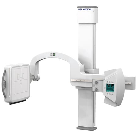
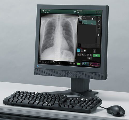
At its core, radiography represents the original form of medical imaging, utilizing radiation to capture images of the body’s internal structure on metal plates. This technique laid the groundwork for more advanced forms of imaging, such as CT scans and MRIs, which offer detailed views of body parts through different technological approaches. While radiography relies on radiation, MRI uses powerful magnetic fields, and CT scans employ computer-processed combinations of many X-ray measurements taken from different angles to produce cross-sectional images.
Navigating the Safety of Medical Imaging
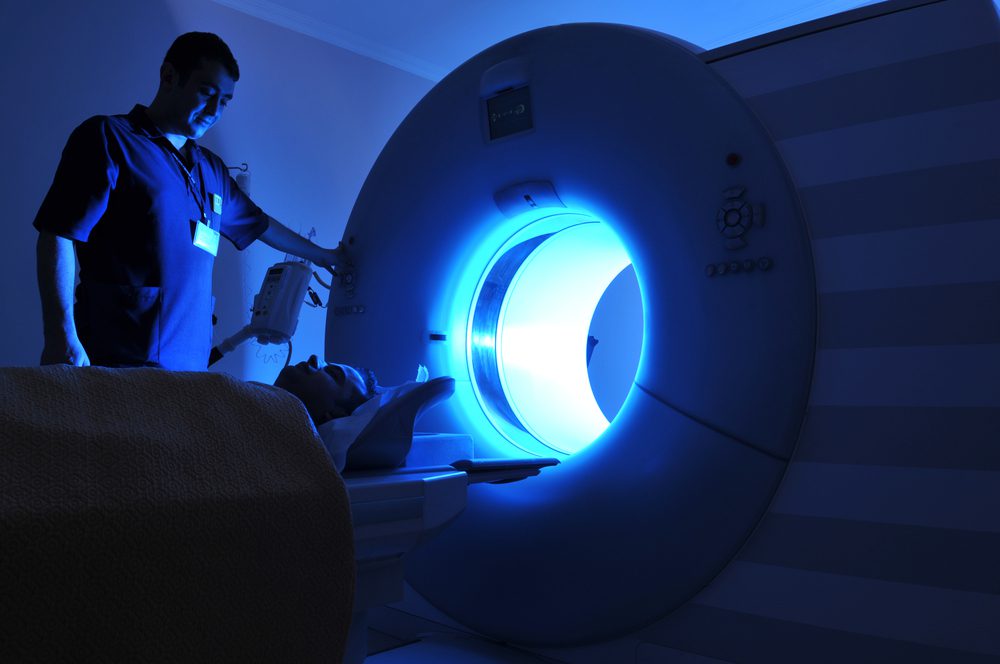
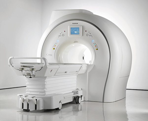
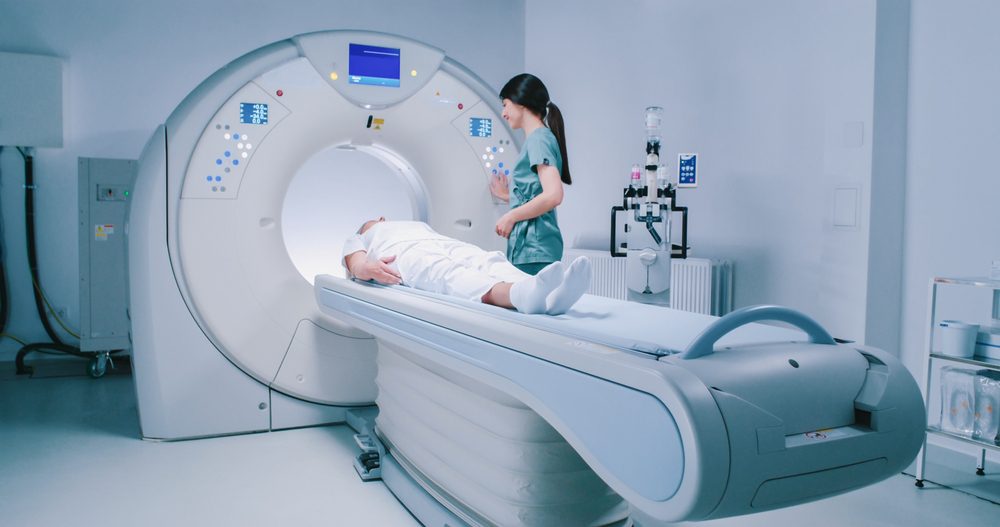
When considering safety, MRI emerges as a frontrunner among medical imaging modalities due to its minimal radiation exposure, making it a preferable option for a wide range of diagnostic needs. However, MRIs are generally advised against for pregnant women due to potential risks to the developing baby. On the other hand, Computer Tomography (CT) provides a rapid and detailed imaging solution, making it a valuable tool in emergency and outpatient care settings. Despite the radiation involved, CT scans are sometimes recommended for pregnant women for their ability to deliver precise images quickly, aiding in specific diagnostic challenges.
Specialized Imaging Techniques
The domain of medical imaging is rich with specialized techniques, such as Vascular Interventional Radiography, which leverages imaging technology to explore and treat blood vessel irregularities. This method plays a pivotal role in managing conditions like heart disease and stroke. Similarly, Sonography uses sound waves to paint images of internal organs and is particularly useful in monitoring pregnancies and detecting conditions like kidney stones early.
Sonography: A Sound Wave Symphony
Sonography stands out for its use of sound waves to generate images, a technique pivotal for examining the heart, lungs, and abdominal organs. It’s indispensable for prenatal care, allowing for the early detection of potential issues in a developing fetus, from gestational diabetes to high blood pressure. Beyond pregnancy, sonography offers a non-invasive method to identify and manage various health conditions, including the early stages of varicose veins and kidney stones, facilitating timely intervention.
The Future Is Bright with Great Lakes Imaging
The field of medical imaging is a testament to the incredible advancements in healthcare technology, providing critical tools for diagnosis and treatment. From the foundational practice of radiography to the sophisticated analysis provided by MRI, CT scans, and sonography, these technologies continue to evolve, driven by a commitment to improving patient care. Great Lakes Imaging remains at the forefront, ready to equip healthcare professionals with the knowledge and tools needed to harness these technologies effectively. Whether you’re integrating new services into your practice or seeking to enhance existing capabilities, we’re here to support your journey toward excellence in patient care.