Sub-Total: $0.00


20/20 Imaging Tether...
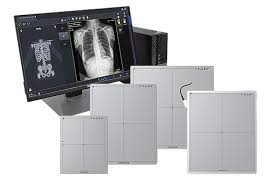
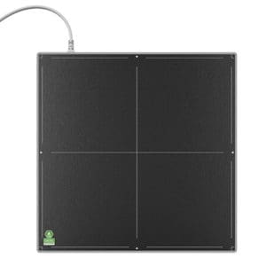
20/20 imagingSPECIFICATIONS
- Image Sensor: a-Si (amorphous Silicon) TFT
- Scintillator Type: FP: CSI (Cesium)
- Pixel Size: 139um
- Pixel Matrix: 3072×3072
- Active Area: 16.81″x16.81″
- Line pair per mm: 3.6 lp/mm
- Dimensions: 18.1×18.1x.59in
- AD conv. 16-bit
Description
Opal-RAD Software
user friendly patient database, imaging tools, & acquisition
Powered by medical imaging’s fastest distribution engine and open Web-based technologies, Opal-RAD will change the way you think about taking x-rays.
A feature-rich, scalable and flexible picture archiving and communication system (PACS), Opal-RAD enables advanced digital image interpretation, management and archiving at a uniquely affordable price.
(mOpal) also available as an add-on
The Diagnostic Image Viewer screen provides a wealth of tools and options to assist in reading and manipulating high-resolution medical images.
- State-of-the-art DICOM viewer delivers powerful, intuitive workstation functionality
- Quick function shortcuts integrated for efficiency
- Full Chiropractic (DC) tools included with package purchase
- Available Chiropractic stitching (Manual & Automatic) contact sales for details
- Podiatric (DPM) Tools available†
- WORLDWIDE access: view from anywhere!*
- High-resolution multi-monitor support
- 4k monitor support
- Customize screen layouts; up to 9 images per monitor
- Fully customizable settings to accommodate your specific needs
- Compare images (post/pre op)
- Refresh (see saved/available images, while study is being performed)
- Custom Toolbox (see top-left of above image), fully customizable annotations/tools
- Bone Enhancement – reprocess images sharper for enhanced diagnosis